x_ray
What is an X-ray Source?
Figure 1: Filament-based X-ray Tube
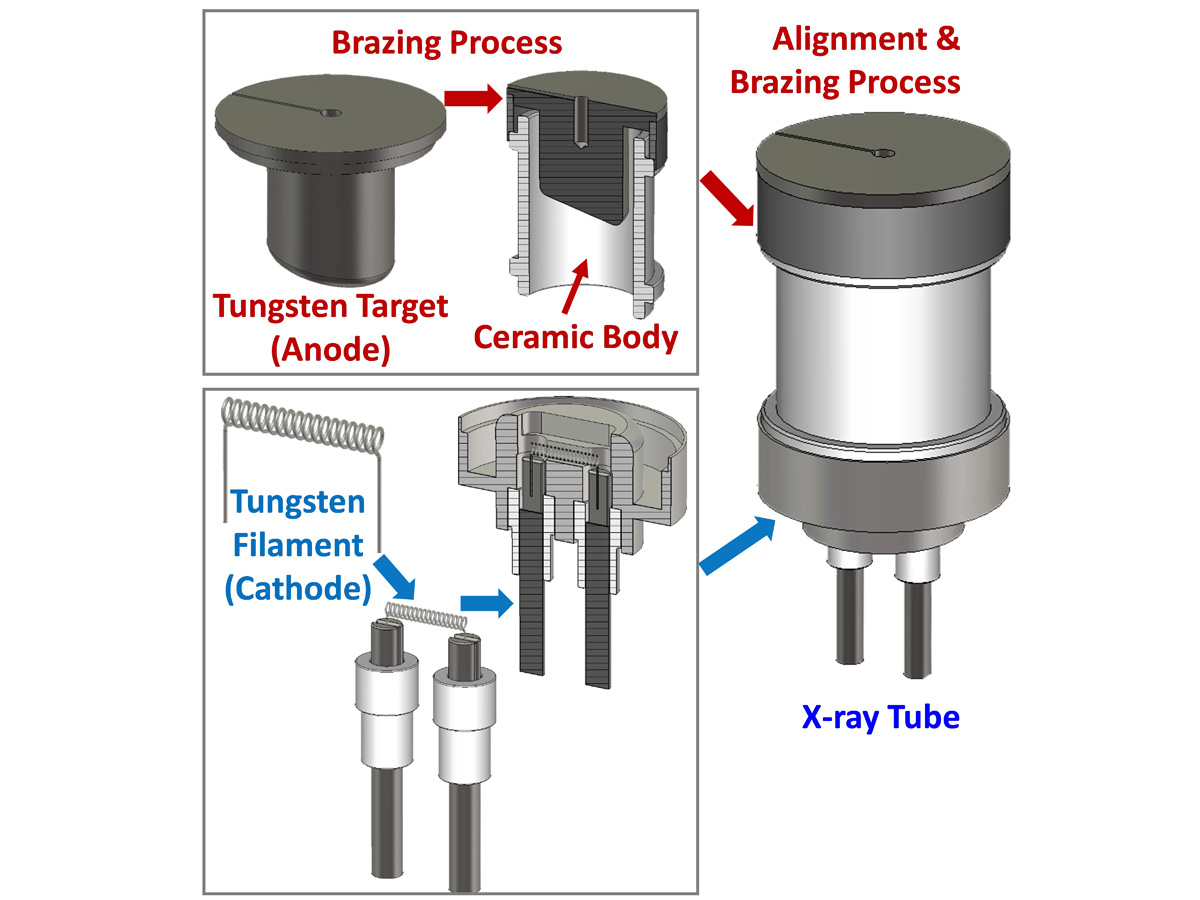 Figure 2: CNT-Based Electron Gun (cathode)
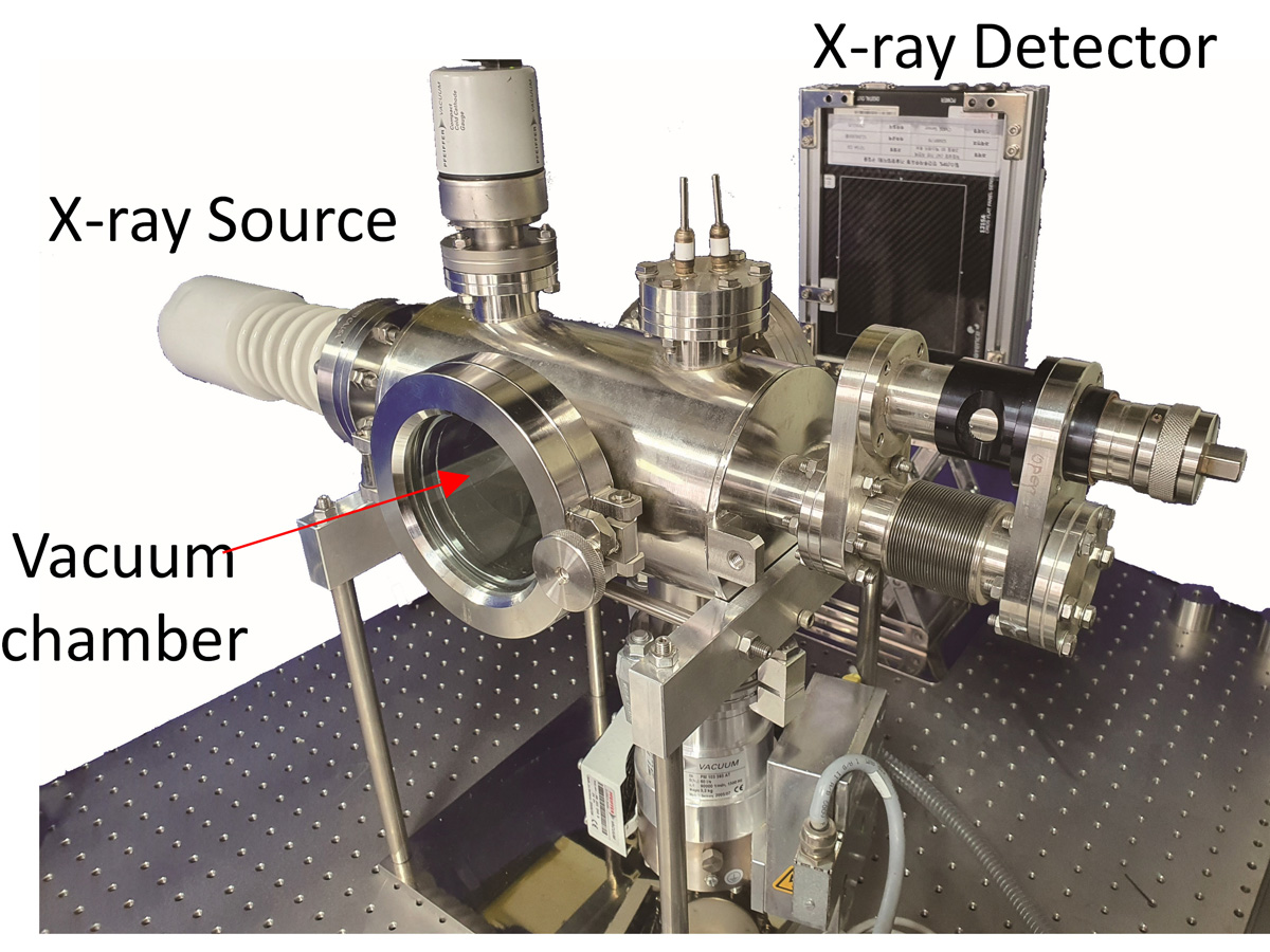
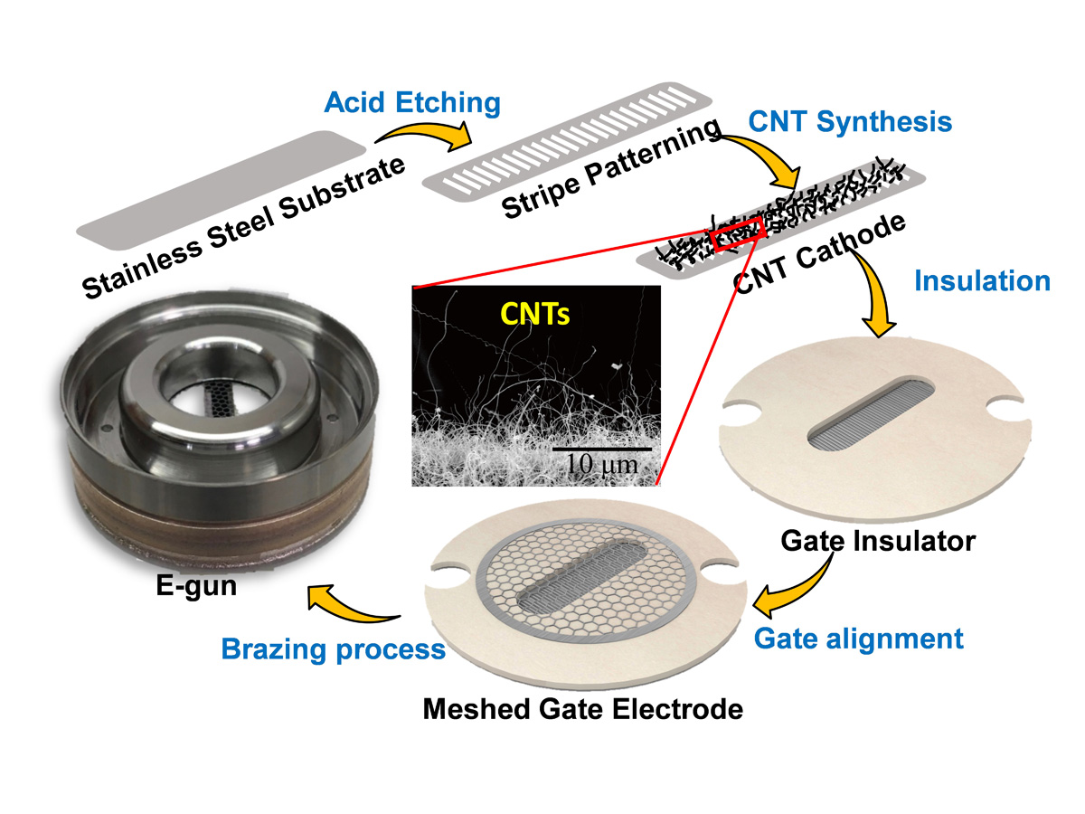 Figure 3: CNT-Based X-ray System
 For over a century now, filament-based thermionic electron-based sources have been the core technology behind all X-ray applications. The filament is typically a tungsten coil encased in a large vacuum glass/ceramic tube (see Figure 1). A negative voltage is applied to the filament, which is the source of electrons and is known as the cathode. The cathode creates free electrons through a process called thermionic emission. Another component found inside the vacuum tube is the anode. The anode absorbs the electrons emitted by the cathode and creates X-rays. When a high amperage current (the mA) is circulated through the filament it gets extremely hot – so hot that it creates a cloud of free electrons. This is the process of thermionic emission. Generating X-rays this way requires time, to pre-heat and cool down the system. As a result, filament-based thermionic X-ray sources can not be turned ON and OFF at fast speeds, which makes them unsuitable for high-end digital security and medical inspection techniques as these demand fast switching X-ray sources.
In MNDL, we have surpassed the limits of the conventional thermionic X-ray systems by replacing the filament-based thermionic electron sources with carbon nanotube (CNT)-based electron sources. CNT-based systems have the potential to fulfil most of the requirements demanded by the high-end digital security and medical inspection techniques. CNTs are cold-cathode field-emitters, meaning that they can be swiftly switched ON and OFF, therefore enabling pulsed emissions. The development of a CNT-based electron gun (cathode) is shown in Figure 2. This electron gun and an anode are inserted into a vacuum chamber (such as the one shown in Figure 3) to generate X-rays. References:
1. Int. J. Imaging Syst. Technol. 31, 3 ,1128, September (2021).
2. IEEE Transactions on Electron Devices, 68, 9, 4705, September (2021).
3. IEEE transactions on Electron Devices, 66, 12, 5301, December (2019).
Figure 2: CNT-Based Electron Gun (cathode)
Figure 3: CNT-Based X-ray System
For over a century now, filament-based thermionic electron-based sources have been the core technology behind all X-ray applications. The filament is typically a tungsten coil encased in a large vacuum glass/ceramic tube (see Figure 1). A negative voltage is applied to the filament, which is the source of electrons and is known as the cathode. The cathode creates free electrons through a process called thermionic emission. Another component found inside the vacuum tube is the anode. The anode absorbs the electrons emitted by the cathode and creates X-rays. When a high amperage current (the mA) is circulated through the filament it gets extremely hot – so hot that it creates a cloud of free electrons. This is the process of thermionic emission. Generating X-rays this way requires time, to pre-heat and cool down the system. As a result, filament-based thermionic X-ray sources can not be turned ON and OFF at fast speeds, which makes them unsuitable for high-end digital security and medical inspection techniques as these demand fast switching X-ray sources.In MNDL, we have surpassed the limits of the conventional thermionic X-ray systems by replacing the filament-based thermionic electron sources with carbon nanotube (CNT)-based electron sources. CNT-based systems have the potential to fulfil most of the requirements demanded by the high-end digital security and medical inspection techniques. CNTs are cold-cathode field-emitters, meaning that they can be swiftly switched ON and OFF, therefore enabling pulsed emissions. The development of a CNT-based electron gun (cathode) is shown in Figure 2. This electron gun and an anode are inserted into a vacuum chamber (such as the one shown in Figure 3) to generate X-rays. References:
1. Int. J. Imaging Syst. Technol. 31, 3 ,1128, September (2021).
2. IEEE Transactions on Electron Devices, 68, 9, 4705, September (2021).
3. IEEE transactions on Electron Devices, 66, 12, 5301, December (2019).